

Outline:
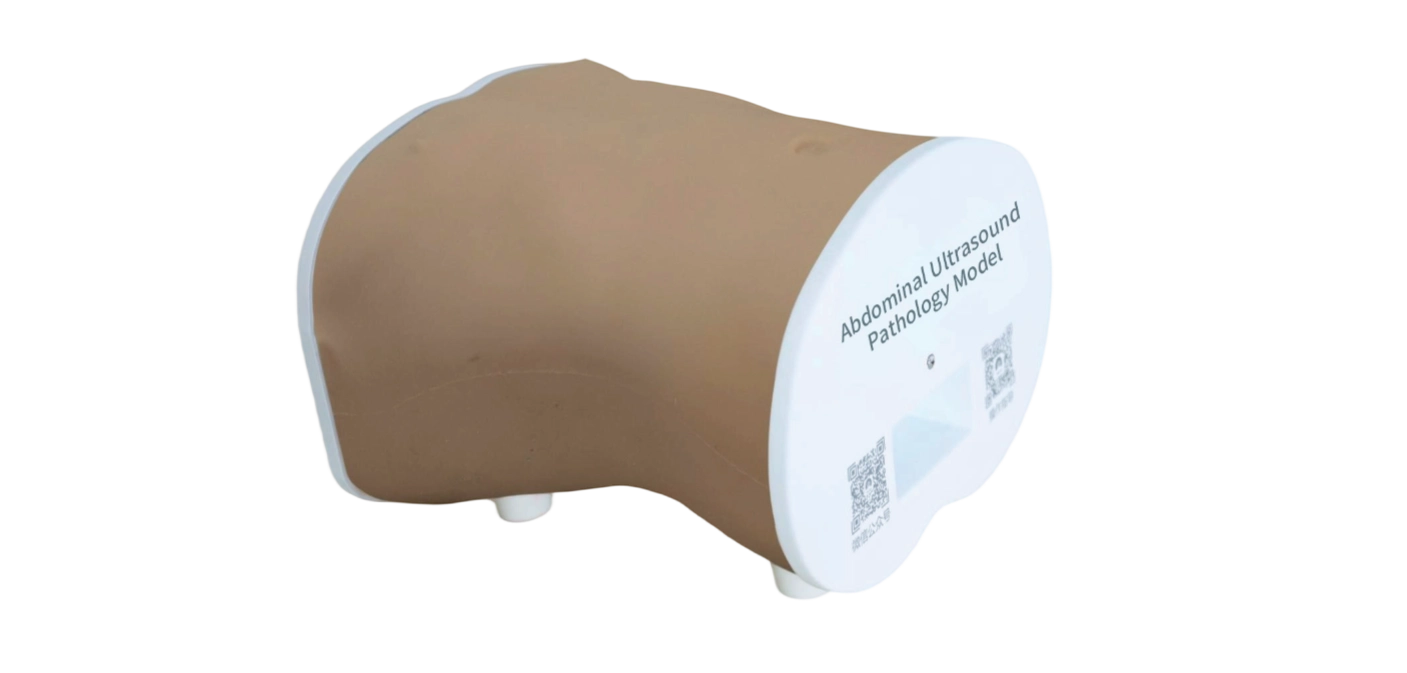
This product is a true-to-life male torso with accurate anatomical landmarks, designed for abdominal ultrasound pathology training. It provides an effective solution for enquiring competencies in routine abdomen examination including abdominal ultrasound techniques, ultrasound-guided biopsy and diagnostic interpretation of pathologic lesions on sonographic imaging. The realism of the product and the skillset specific to abdominal examination techniques will bring learners competence and confidence.
Skills Gained
• Abdominal ultrasound scanning techniques
• Organ scanning
• Standard imaging planes
• Lesion identification and measurement
• Ultrasound-guided liver and kidney puncture biopsy
Features
Realism
1) Compatible with clinical ultrasound machines to acquire lifelike diagnostic images during training.
2) The sonographic imaging demonstrates accurate organ sizing, true-to-life acoustic properties including organ dimensions, density, speed of sound propagation, attenuation, and echogenicity.
Anatomy
1) Transparent abdominal model of an adult male, ranging from the 6th rib to the umbilicus
2) Surface landmarks include the costal margin, xiphoid process, and umbilicus, with major abdominal organs and common pathological lesions.
3) Bionic skeletal structures include ribs and sternum.
4) The model contains 12 anatomical structures, including the diaphragm, esophagus, stomach, duodenum, jejunum, liver, gallbladder, pancreas, spleen, kidneys, adrenal glands, and ureters.
Key features
This product enables imaging of normal tissue structures of each individual organ as well as digestive system, and common pathologies of liver, gallbladder, kidneys, pancreas and spleen.
Liver:
1)The liver has 6 anatomical structures, including left lobe, right lobe, caudate lobe, ligamentum teres hepatis, first hepatic portal, second hepatic portal, etc., containing the portal venous system and hepatic veins, with an intact liver capsule
2)Lesions:
· Hepatic parenchyma was visualized in ultrasound examination. Sonography demonstrates hepatic space-occupying lesion.
· Space-occupying lesions: hyperechoic, hypoechoic, isoechoic, and anechoic cystic lesions.
· Lesion morphology: oval, round, and irregular. The irregular lesions appeared hypoechoic, contained calcific foci, and exhibited ill-defined margins.
3)Ultrasound-guided liver biopsy is supported.
4)The liver can be divided into four sectors via the left, right, and middle hepatic veins during the ultrasound examination. At the transverse plane, the horizontal sections of the right and left portal vein branches present as a linear or band-like transverse fissure structure, further subdividing the hepatic sectors into eight segments according to the Couinaud Nomenclature.
Gallbladder
1) Gallbladder contains 7 anatomical structures, including cystic duct, common bile duct, left hepatic duct, right hepatic duct, cystic neck, cystic body, and cystic fundus.
2) Lesions: gallstones and polyps, during the ultrasound examination, gallstones appear as hyperechoic with posterior acoustic shadowing.
Kidneys
1) Both kidneys are anatomically intact and bean-shaped.
Bilateral renal regions can be examined via transverse and longitudinal plane ultrasound scanning in the left and right upper abdominal quadrants, as well as through dorsal and flank approaches, which allows for the visualization of 7 structures, including renal capsule, renal cortex, renal medulla (renal papilla, renal pyramid, renal column), renal pelvis, major calyces, minor calyces, and ureter.
2) Lesions:
· 2 renal calculi lesions at the major calyces and renal pelvis, presenting hyperechoic echogenicity with posterior acoustic shadowing.
· 1 renal tumor lesion at the upper pole of the left kidney, with indistinct margin, presenting hyperechoic echogenicity without posterior acoustic shadowing and internal echotexture is homogeneous.
· 1 renal tumor lesion at the upper pole of the right kidney, presenting isoechoic echogenicity with clear margin and internal echotexture is heterogeneous, containing hyperechoic calcified foci.
3) Ultrasound-guided renal biopsy is supported.
4) The adrenal glands can be detected superior to the bilateral renal areas.
Pancreas:
1) The pancreas contains the pancreatic duct. A well-defined margin with homogeneous internal echotexture from the pancreatic parenchyma and a slender pancreatic duct can be visualized at the upper abdomen.
2) Lesions: When scanning the pancreatic head, a solid hypoechoic mass with clear margin is visualized, consistent with characteristic imaging features of a pancreatic space-occupying lesion.
Spleen:
1)The spleen contains splenic hilum. The splenic capsule and splenic parenchyma can be visualized during ultrasonographic scanning along the anterior and posterior axillary lines.
2) Lesions:
· 1 splenic cyst lesion demonstrates a circular anechoic area with a smooth-walled capsule on ultrasonography.
· 1 splenic tumor that demonstrates an ovoid hyperechoic mass with clear margin.
Digestive system
The model incorporates 11 anatomical structures, including esophagus, cardia, gastric body, lesser curvature, greater curvature, gastric antrum, pylorus, duodenal bulb, descending duodenum, horizontal duodenum, and adjacent pancreatic, which exhibit characteristic contrast-enhanced ultrasound imaging features during ultrasound examination.
Build-in blood vessel
1) The model contains 24 vessels, including inferior vena cava, abdominal aorta, celiac axis, common hepatic artery, proper hepatic artery, portal vein, right intrahepatic portal vein branch, left intrahepatic portal vein branch, interlobar vein, hepatic vein, left hepatic vein, right hepatic vein, middle hepatic vein, superior mesenteric artery, superior mesenteric vein, inferior mesenteric artery, inferior mesenteric vein, splenic artery, splenic vein, left renal artery, left renal vein, right renal artery, right renal vein, left testicular artery, right testicular artery.
2) When pressure is applied with an ultrasound probe, venous vessels can be compressed flat, while arterial vessels maintain the shape. True-to-life vessel course and lumen diameter are visualized during the ultrasound examination, with simulated blood inside, which allows for the measurement of vessel length and internal diameter.
Stock code :833047
Address:2nd & 3rd Floor, West 6th Building, 18 West HaiTai Road, Tianjin, China
Postcode:300384
Phone:4006-355-510
+86-22-83711066
Fax:+86-22-83711065
Email:info@tellyes.com

 Ultrasound